
Heart rhythm problems, also called arrhythmias, mean that your heartbeat is too fast, too slow, or irregular.
Your heart has a built-in electrical system that organizes and times your heartbeat. A network of very special cells in your heart muscle conducts electricity — and that electrical signal makes your heart muscle contract.
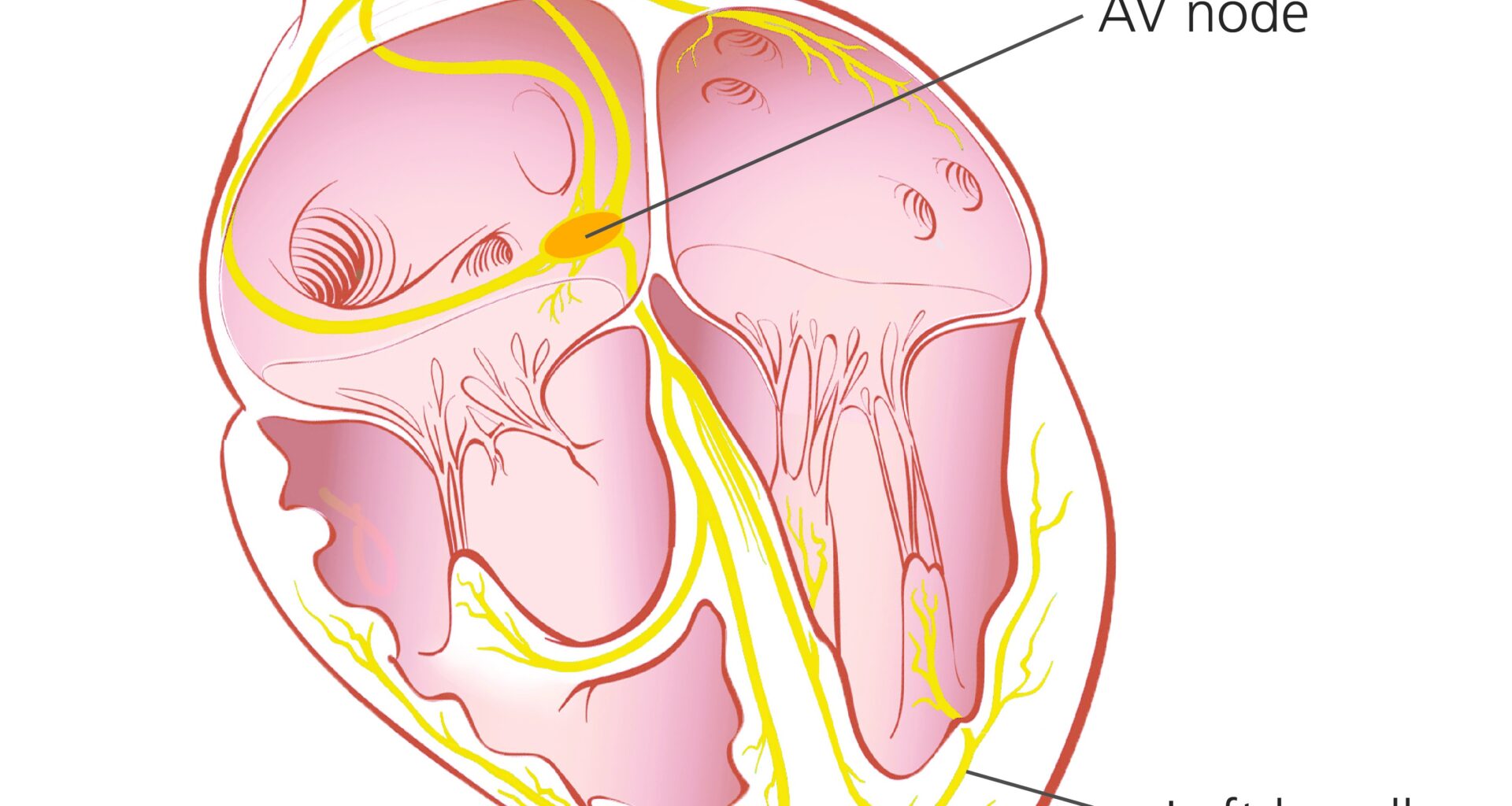
In a healthy heart, the heartbeat starts at a bundle of cells called the sino-atrial (SA) node. The SA node sends an electrical impulse to the upper chambers of your heart, the atria, causing them to contract. Then the signal arrives at another bundle of cells called the atrio-ventricular (AV) node.
The AV node passes the signal to the lower chambers of your heart, called the ventricles, causing them to contract as well. The process repeats with every heartbeat and is highlighted in the image below in yellow.
Problems happen when the SA node, also called the heart’s natural pacemaker, is no longer in control of the electrical impulses in your heart muscle. This disrupts your heart’s normal rhythm and makes it work less efficiently.
Abstract
References
- , . Atrial cardiomyopathy an orphan disease or common disorder? Circ Cardiovasc Genet. 2013; 6(1): 5– 6.
- CrossrefPubMedGoogle Scholar
- , , , , , , et al. Isolated atrial amyloidosis suspected by electrophysiological voltage mapping and diagnosed by 99m Tc-DPD scintigraphy. ESC Heart Fail. 2020; 7(6): 4305– 10.
- Wiley Online LibraryPubMedWeb of Science®Google Scholar
- , , , , , , et al. Accuracy of left atrial fibrosis detection with cardiac magnetic resonance: correlation of late gadolinium enhancement with endocardial voltage and conduction velocity. Europace. 2021; 23(3): 380– 8.
- CrossrefPubMedGoogle Scholar
- , , , , , , et al. EHRA/HRS/APHRS/SOLAECE expert consensus on atrial cardiomyopathies: definition, characterization, and clinical implication. Europace. 2016; 18(10): 1455– 90.
- CrossrefPubMedWeb of Science®Google Scholar
- , , , . Left atrial voltage mapping: defining and targeting the atrial fibrillation substrate. J Interv Card Electrophysiol. 2019; 56(3): 213– 27.
- CrossrefPubMedWeb of Science®Google Scholar